Early Detection of Skin Cancer
Read our Facial basal cell carcinoma publication in the BMJ (free)
I specialise in the early detection of skin cancers such as basal cell carcinoma (BCC), squamous cell carcinoma (SCC) and malignant melanoma (MM) and pre-cancers of the skin such as Bowen’s disease (IEC) and actinic keratoses.
The early detection of skin cancer is crucial to improving outcome – especially for MM and certain SCC. The sooner a MM is picked up and excised the better the chances of long term non-recurrence (“cure”).
The best way to making an early diagnosis is through a thorough history of the problem, and close examination of the lesion.
Dermatoscopy helps a great deal with diagnosis. Using a dermatoscope can take years of experience to look at lesions to decide which are benign, pre-cancerous, suspicious or cancerous lesion.
 |
|
Dermatoscope
|
|
Click here for a dermatoscopy resource
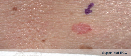
Below are some examples of early diagnosis of skin lesions.
For example, there are 2 examples below which show small, early, non-descript skin lumps. These could have been overlooked. Dermatoscopically, they show the hallmark features (arborising telangiectasia, blue grey ovoid globule) of basal cell carcinoma. Surgery was performed which confirmed complete excision of BCC.
Lesion on forehead, difficult to diagnose visually
 |
 |
Small flesh toned skin lump -
hard to see
|
Hard to diagnose |
 |
 |
Dermatoscopy reveals arbrising
telangiectasia - thus a bascal
cell carcinoma
|
Non-descript skin lump on back |
 |
|
Dermatoscopy shows BCC
|
|
The next two photos show a lesion which was suspected of being a cancer but which is completely harmless.
 |
 |
Small skin lesion - could this be
a cancer
|
Dermatoscopy shows hairpin
veesels - this is a harmless
seborrheic keratosis dermatoscopy |
CompoundNaevus (Normal Mole)
 |
 |
|
Dermatosopy shows normal mole |
Atypical Naevus (irregluar mole)
 |
 |
Irregular mole
|
Dermatoscopy + exision show
irregular mole |
Malignant Melanoma
 |
 |
15 mm irregular mole
|
Dermatoscopy = malignant melanoma
(MM), excision confirms |
Click here for
Dermatoscopy atlas 1
Dermatoscopy atlas 2
Red dot basal cell carcinoma: an early & distinct clinical presentation.
|